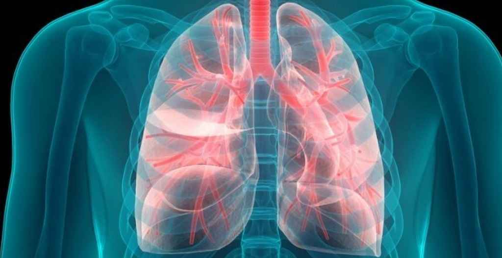
Lung nodules are focal, round, or oval dense opacities in the lung, with well-defined or indistinct borders and a diameter ≤30 mm. They may be solitary or multiple and typically lack associated atelectasis, hilar lymphadenopathy, or pleural effusion.
Lung nodules are classified by:
Etiology
Lung nodules can be divided into benign nodules and malignant nodules.
- Benign nodules
Benign tumors, infections, rheumatologic disorders, congenital anomalies, pulmonary hemorrhage.
- Malignant nodules
Bronchogenic carcinoma (pre-invasive/invasive lesions), lymphoma, sarcoma, pulmonary metastases.
Density
A round/oval lesion with sufficient density to obscure underlying vessels and bronchi.
- Subsolid nodule
A round/oval lesion with hazy density that does not obscure blood vessels and bronchi, appearing as ground glass. For this reason, experts call it a ground-glass nodule/opacity (GGN/GGO).
Sub-solid lung nodules fall into two categories: pure ground-glass nodules (pGGNs) and mixed ground-glass nodules (mGGNs)—also known as part-solid nodules (PSNs).
Sub-solid nodules that are potentially malignant or malignant are typically linked to lung adenocarcinoma. This can range from atypical adenomatous hyperplasia (AAH) to adenocarcinoma in situ (AIS), then to microinvasive lung adenocarcinoma (MIA), and finally to invasive adenocarcinoma (IA).
Size
- Very small nodule
<5 mm in diameter (volume <100 mm³)
- Small nodule
5-10 mm in diameter, (volume 100-300 mm³)
- Standard nodule
11-30 mm in diameter (volume ≥300 mm³)
Number
- Solitary nodule
Single lesion
- Multiple nodules
2 or more lesions
Summary
Accurate classification guides clinical management. Understanding nodule characteristics enables optimal patient care.
